New technique for treating spinal dislocation involving nerve compression: Full Endoscopic TLIF (Transforaminal Lumbar Interbody Fusion) reduces incision size and achieves good outcomes.
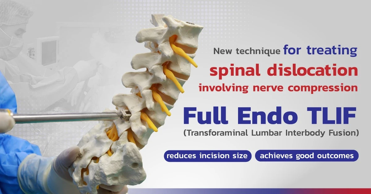
“The treatment of spinal dislocation involving nerve compression, or spinal bone impingement, using the TLIF (Transforaminal Lumbar Interbody Fusion) technique is a procedure that involves connecting the vertebrae to prevent movement of the patient’s spine and replacing worn-out spinal cushions with artificial ones. In the past, this procedure was performed through open surgery. However, there have been advancements in the form of MIS TLIF, which involves inserting a tube and dissecting some muscle tissue before placing the artificial cushion. Most recently, a new technology called Full Endo TLIF (Full Endoscopic TLIF) has been developed for treating spinal dislocation involving nerve compression. This technique uses an endoscope to insert the artificial cushion, resulting in minimal muscle disruption, improved safety compared to traditional methods, reduced incision size, and better outcomes.” – Dr. Methee Phakkawet, Spine Specialist Physician.
Spondylolisthesis, also known as “vertebral slippage” or “slipped vertebra,” occurs when one of the vertebrae in the spine moves forward from its normal position. The vertebrae in the spine are capable of movement, but this condition typically occurs in the lower back, specifically at the L4-L5 level. It is more commonly found in individuals aged 50 and above and is more prevalent in females than males. This is because women generally have weaker muscle strength and less robust ligaments than men, making them more susceptible to this condition. Research conducted by the National Institute of Health in the United States has provided comparative data between the treatment methods of Endoscopic TLIF and MIS TLIF. It has been found that within a six-month period, patients treated with Endoscopic TLIF experience better recovery. This aligns with the data from patients who received treatment for spondylolisthesis involving nerve compression using the Full Endo TLIF technique at the S-Spine and Nerve Hospital, where this technique has been implemented since 2023. In just four months, more than 20 patients have undergone this treatment, and each case has yielded satisfactory results.
The treatment method for spinal dislocation involves nerve compression using the Full Endo TLIF technique.
Currently, there are various techniques available for treating spinal dislocation, and S-Spine and Nerve Hospital has introduced a new method that minimally affects the patient’s muscle. It is called the Full Endo TLIF (Full Endoscopic TLIF) technique, which involves using a minimally invasive approach with an endoscope instead of making a large incision. The endoscope is inserted without the need to cut through muscle tissue. The surgeon will remove the compressed nerve structures, such as the cushion, nerves, and problematic parts of the vertebrae. Afterward, an artificial spinal cushion made of PEEK (Polyether Ether Ketone) is inserted through the endoscope. Interbody fusion is performed using percutaneous screws, adding three additional screws. It can be observed that this technique minimally disrupts the spinal structure, and the postoperative pain of the patients is significantly reduced compared to the traditional treatment methods.
Regarding Endoscopic TLIF, there are two approaches. The first approach is the two-hole technique commonly known as the Endoscopic assist TLIF, which is commonly used in most hospitals in Thailand. Although it involves the use of an endoscope, it is similar to open surgery as it requires instruments to be inserted into both sides of the body, passing through various layers of muscle tissue.
When the camera and instruments are maneuvered to find the required angle or position for treatment, it can cause more muscle and nerve tissue trauma. Additionally, the camera and instruments are not located in the same place, which increases the risk of errors when using the instruments. This procedure takes approximately two and a half hours to perform.
The second approach is the single-hole technique, or Full Endo TLIF (Full Endoscopic TLIF). In this method, a specialized camera with a channel for inserting instruments is used for treatment. It involves removing the problematic cushion between the vertebrae and the ability to replace it with an artificial cushion. The instruments do not directly damage the muscles, resulting in less trauma compared to the two-hole technique. The surgeon can constantly visualize the instruments during the procedure, and the treatment time is approximately two hours. After the treatment, patients recover quickly, the incision is small, and within one week, they can walk comfortably. It’s worth noting that this Full Endo TLIF technique is only available at the S-Spine and Nerve Hospital.
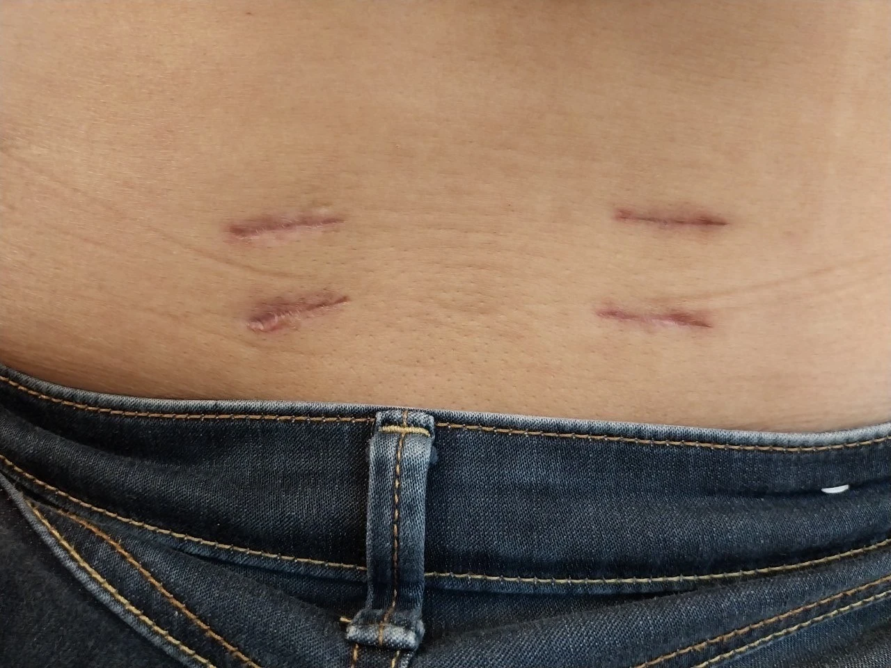
After 1 week surgery wound technique with Full EndoTLIF
How does the treatment technique of Full Endo TLIF differ from other techniques?
Full Endo TLIF or spinal fusion surgery performed using an endoscope, known as Endoscopic TLIF, involves using an endoscope to access the spine without cutting significant portions of the surrounding muscles. This approach minimizes blood loss and results in small incisions, typically no larger than 1 centimeter, equivalent to the size of a screw that holds the vertebrae together. Patients who undergo this surgery can usually get up and walk shortly after the procedure. The post-operative back pain is significantly reduced, and patients may require minimal or no pain medication. The hospital stay typically lasts around 2–4 days, after which patients can resume their normal daily activities at home. This technique, Full Endo TLIF, is effective in treating spondylolisthesis or spinal instability. However, it requires a skilled and experienced surgeon to ensure safety and successful outcomes.
MIS TLIF (Minimally Invasive Transforaminal Lumbar Interbody Fusion) or the treatment of spinal fusion to stabilize the affected nerves is widely used nowadays. The incision size is smaller compared to open surgery, typically around 3 centimeters in the middle of the back, to insert the instruments into the problematic vertebrae and remove a small portion of the muscle. However, it is not as extensive as open surgery. Before the surgeon changes the intervertebral disc and inserts four screws, each with an incision size not exceeding 1 centimeter, equivalent to the size of the screw that holds the vertebrae, it aims to enhance stability for the spine. Within the first 24 hours after the surgery, pain medication is required, but there is no need for drainage tubes. Additionally, patients recover faster compared to those who undergo open surgery. They typically stay in the hospital for only 3–4 days and can then return home.
Open surgery to stabilize and treat spinal vertebrae displacement by fixing screws is not commonly preferred nowadays due to the high risks involved. This method involves making an incision in the mid-back region, where the surgeon will dissect muscles and remove a portion of the vertebrae. Metal rods and screws are then inserted from the back to stabilize the spinal vertebrae and fuse them together. Additionally, a drainage tube is placed to allow blood to drain. Typically, within 24 hours after the surgery, the patient needs to remain still in bed as the incision is large, approximately 5–7 centimeters, depending on the size of the affected vertebrae. The patient usually stays in the hospital for 4–5 days until the incision pain subsides. Physical therapy is necessary afterward to prevent muscle weakness or stiffness, and it may take approximately 2–3 months for the patient to return to normal walking abilities.
Best practices for treating spinal vertebrae displacement and nerve compression using the Full Endoscopic TLIF technique.
After treatment, most patients will experience improvement and can resume their normal work routine. However, in some cases, it may take approximately 4–8 weeks before they can return to work. This duration depends on the patient’s overall physical strength. Even though patients may feel better after surgery, if they continue to engage in activities such as bending, sitting on the floor, squatting, or lifting heavy objects, there is a chance that they may require another surgery in the future.
During the recovery period, patients can start exercising by walking for about 5–10 minutes during the first 6 weeks. This is because the muscles and nerves may still need time to fully recover. However, after 6 weeks, patients can gradually increase the distance they walk or the time they spend walking, as the internal wound improves. Engaging in such exercises will aid in a quicker return to normal physical function.
Taking care of ourselves to prevent of spinal dislocation involving nerve compression, or spinal bone impingement, is crucial because prevention is always better than correction. Therefore, we should prioritize maintaining our spinal health in the long term. One effective way is to strengthen our core muscles, the central muscles of the torso. Before engaging in exercise or sports activities, it is important to stretch our muscles to prepare them for the activity. Limiting the duration of activities that put excessive strain on the spine, especially those involving twisting movements or heavy lifting, can help reduce the risk of spinal misalignment. Additionally, it is advisable to maintain healthy body weight as it contributes to minimizing the risk of spinal misalignment.
Reference:
Dr. Matee Phakawech, Spine Specialist Physician.
Percutaneous Endoscopic Transforaminal Lumbar Interbody Fusion: Technique Note and Comparison of Early Outcomes with Minimally Invasive Transforaminal Lumbar Interbody Fusion for Lumbar Spondylolisthesis https://www.ncbi.nlm.nih.gov/pmc/articles/PMC7910530/


